

BREEDER
QUALIFICATIONS:
KENNEL NAME: Haus Merkel
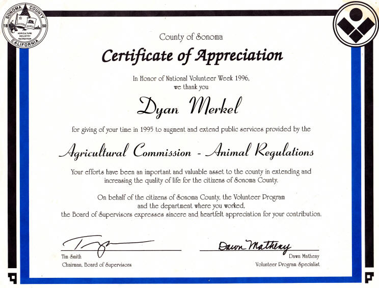
Breeder Name: Dyan Merkel
Website: http://www.hausmerkel.com
Location: North Texas
Contact Info: vhmerkel@yahoo.com
214-755-5755
KENNEL NAME: Haus Merkel
Breeder Name: Dyan Merkel
Website: http://www.hausmerkel.com
Location: North Texas
Contact Info: vhmerkel@yahoo.com
214-755-5755
HEALTH TESTS:
Hip/Elbow
Certifications: Yes
Degenerative Myopathy
Certifications: Yes
Thyroid tests: Yes
CERF tests: No
TRAINING:
Titles/certifies
breeding stock in
discipline?: Yes
BREEDING STOCK:
Raises breeding
stock from
puppies:
Yes
Titles dogs bred on
premise: Yes
Imports titled
breeding stock: Yes
Buys from other
Breeders: NO
MORE!
SCHUTZHUND/IPO:
Has trained in
Schutzhund: Yes
HOT from puppy to
SchH3: Yes
HOT and bred to
SchH3: Yes
CONFORMATION/BREED
SURVEY:
Show ratings: Yes
Breed
survey: Yes
BREEDER
ACCOMPLISHMENTS:
V Putz vom Haus
Merkel SchH3, Kkl 1a
V Ulla vom Haus
Merkel SchH3, Kkl 1a
V Zessa vom Haus
Merkel SchH2, IPO3,
Kkl1a
V Riesa vom Haus
Merkel SchH2, Kkl 1a
V Puma vom Haus
Merkel SchH2, Kkl 1a
V Emma vom Haus
Merkel SchH1, Kkl 1a
SG Wickie vom Haus
Merkel IPO1 a
SG Vessa vom Haus
Merkel IPO1 a
SG1 Clar vom Haus
Merkel SchH1 a
Ch Merkel's Opium
SchH1
V Merkel's Arletta
SchH1, a
SG, VP2 Nixe vom Haus
Merkel BH, AD, a
SG Feli vom Haus
Merkel BH, AD, a
Ch Merkel's Leica
UDT, OFA
Merkel's Leibchen
Shiloh UDT, OFA
Ch Merkels Sangria
UDT, OFA
United States
Grand Victrix
Ch
Merkels Vendetta ROM, OFA
Ch Merkel' Quaestor
CD, ROM, OFA
Ch Merkel's Essence
UD, OFA
Merkel's Coda vom
Jennerick CDX, OFA
National
Certified Search &
Rescue
Lieb vom Haus Merkel
OFA,
National
Obedience
Winner
Ch Merkel's Cut Up
of Timmee UDT, OFA
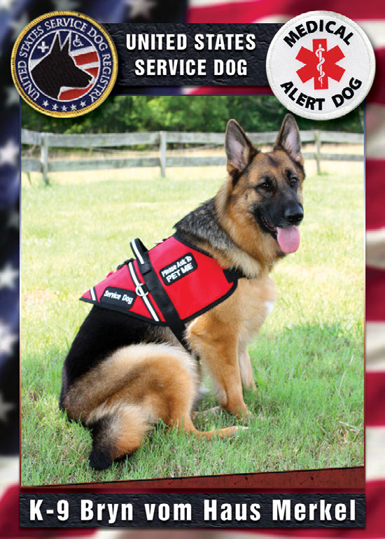
Certified United
States
Service Dog
Bryn vom Haus Merkel
CGC, OFA
Certified United
States
Service Dog
Fred vom Haus
Merkel
Ch Merkel's The
Cutting Edge OFA
Ch Merkel's Virtual
Reality OFA
Ch Merkel's Tequila
CD, OFA
Ch Merkel's Sante Fe
OFA
Merkel's Spellbound
ROM,OFA
Merkel's Emma ROM ,
OFA
Merkel's Estes CD,
near ROM
2009 Annual Achievement Award Recipient
Ch Merkel's Heart's are Wild
CDTDTC HIC CGC
TDI
OFA
Ch Merkel's Heart to
Heart CD, OFA
PRODOMINANT LINES
USED:
World Sieger Larus von Batu
SchH3 Kkl 1a
World Sieger Zamp vom Thermodos
SchH3, Kkl 1
World Sieger Yasko vom
Farbenspiel SchH3,Kkl 1a
VA Dux della
Valcuvia SchH3,
Kkl1a
Kirschental
WRITTEN GUARANTEE:
Yes
EDUCATION
AVAILABLE:
Yes
LIFETIME
SUPPORT:
Yes
YEARS AS BREEDER:
46